
The humerus is the longest bone in the upper limb, connecting the shoulder and elbow joints. It forms the structural foundation of the arm.
1.1 Definition and Overview
The humerus is the longest bone in the upper limb, serving as the structural backbone of the arm. It is a long bone with a distinct proximal end, a cylindrical shaft, and a distal end. Its role is to connect the shoulder and elbow joints, providing a foundation for movement and stability. The humerus is divided into three main regions: the proximal end, shaft, and distal end, each with unique anatomical features. It plays a crucial role in enabling a wide range of arm movements, from flexion to rotation, while supporting the body’s weight and facilitating daily activities.
1.2 Importance in Upper Limb Anatomy
The humerus is vital for upper limb function, serving as the primary bone connecting the shoulder and elbow. It provides attachment points for major muscles, enabling movement and stability. Its structure supports the rotator cuff, allowing shoulder mobility, while its distal end facilitates elbow joint articulation. The humerus also houses nerve pathways and blood vessels essential for limb function. Fractures or injuries to this bone can significantly impair arm movement, highlighting its critical role in anatomy and functionality. Its unique anatomy ensures efficient load-bearing and dynamic movement capabilities, making it indispensable for daily activities and overall upper limb mechanics.
1.3 Brief History of Humerus Bone Studies
The study of the humerus bone dates back to ancient anatomy, with early descriptions by Galen and Vesalius. Modern research expanded with detailed anatomical studies in the 20th century, focusing on its structure and function. Recent advancements include minimally invasive surgical techniques for fractures and implantations, as documented in 2020 studies. Imaging technologies have enhanced understanding of its anatomical landmarks, while clinical cases, like tumor-like cysts post-glucocorticosteroid injections, highlight its complexity. Historical studies have laid the foundation for contemporary medical applications, emphasizing the humerus’s role in upper limb anatomy and its significance in clinical treatments and anatomical education.
Structure of the Humerus Bone
The humerus bone is divided into three main parts: the proximal end, shaft (diaphysis), and distal end, each serving distinct functional roles in upper limb structure.
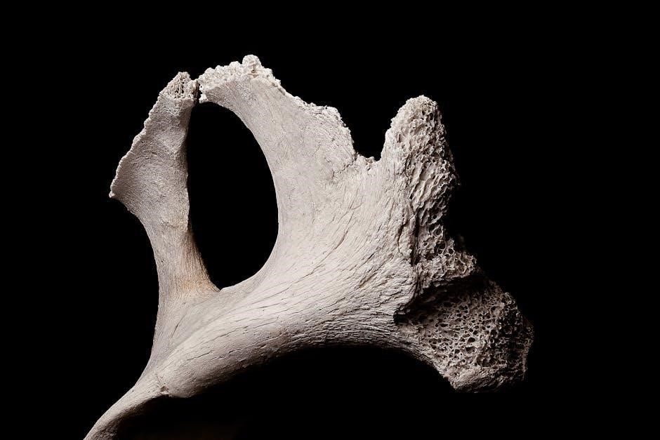
2.1 Proximal End
The proximal end of the humerus is a complex structure designed for articulation and muscle attachment. It includes the humeral head, anatomical neck, and greater and lesser tubercles. These features allow for shoulder joint movement and provide attachment points for the rotator cuff muscles, which stabilize the shoulder. The humeral head articulates with the glenoid cavity of the scapula, enabling a wide range of motion. The anatomical neck separates the head from the tubercles, while the greater and lesser tubercles serve as sites for muscle insertion, facilitating arm rotation and abduction. This region is crucial for upper limb mobility and stability.
2.2 Shaft (Diaphysis)
The shaft, or diaphysis, of the humerus is a long, cylindrical structure that connects the proximal and distal ends. It is triangular in cross-section, providing strength and stability. Along its length, the shaft features the deltoid tuberosity, a roughened area for muscle attachment, and the radial groove, which houses the radial nerve. These anatomical features facilitate movement and sensation in the arm. The shaft’s design allows for efficient transmission of forces from the shoulder to the elbow, enabling a wide range of upper limb movements. Its structure is vital for both mobility and functional support.
2.3 Distal End
The distal end of the humerus is a complex structure that articulates with the radius and ulna bones at the elbow joint. It features two condyles, the capitellum and the trochlea, which form the humeroradial and humeroulnar joints. The capitellum articulates with the radial head, while the trochlea connects with the ulna. The olecranon and coronoid processes of the ulna fit into the olecranon and coronoid fossae of the humerus during flexion and extension. The distal end also includes the medial and lateral epicondyles, which serve as muscle attachment points, enhancing forearm movement and stability. This region is crucial for elbow function and mobility.

Anatomical Landmarks
The humerus features key landmarks, including the humeral head, anatomical neck, greater and lesser tubercles, intertubercular groove, and deltoid tuberosity, each serving specific functional roles.
3.1 Humeral Head
The humeral head is the rounded, proximal end of the humerus, covered by articular cartilage. It articulates with the glenoid cavity of the scapula, forming the shoulder joint. This structure allows for a wide range of motion, including flexion, extension, abduction, and rotation. The head is surrounded by the anatomical neck, a groove separating it from the greater and lesser tubercles. This anatomical design provides a stable yet mobile connection between the arm and shoulder, enabling complex movements essential for daily activities and sports. The humeral head’s shape and articulation make it a critical component of upper limb anatomy.
3.2 Anatomical Neck
The anatomical neck is a slight constriction located distal to the humeral head, marking the transition to the greater and lesser tubercles. It is an important anatomical landmark, often used in surgical procedures like shoulder arthroplasty. The anatomical neck serves as an attachment point for the joint capsule and surrounding ligaments, providing stability to the shoulder joint. Its position and structure are crucial for proper muscle and tendon attachments, ensuring a wide range of shoulder movements. This area is also a common site for fractures, highlighting its clinical significance in orthopedic treatments and upper limb injuries.
3.3 Greater and Lesser Tubercles
The greater and lesser tubercles are prominent bony projections on the proximal humerus, located lateral and anterior to the anatomical neck. These tubercles serve as attachment points for the rotator cuff muscles: the supraspinatus, infraspinatus, teres minor, and subscapularis. The greater tubercle is larger and situated on the lateral aspect, while the lesser tubercle is smaller and positioned anteriorly; These structures are essential for shoulder movement and stability, enabling actions like abduction, rotation, and adduction. Injuries to these areas, such as fractures or tendinitis, are common, particularly in active individuals, emphasizing their clinical significance in orthopedic care.
3.4 Intertubercular Groove
The intertubercular groove, also known as the bicipital groove, is a longitudinal channel on the anterior aspect of the humerus. It lies between the greater and lesser tubercles, extending distally along the shaft. This groove houses the long head of the biceps brachii tendon, providing a protected pathway for tendon movement. The groove is lined with a fibrous sheath, reducing friction during arm movements. Its bony walls also serve as attachments for surrounding muscles and ligaments. This anatomical feature is clinically significant in shoulder injuries and surgeries, particularly those involving the biceps tendon or rotator cuff. It plays a crucial role in shoulder mobility and stability.
3.5 Deltoid Tuberosity
The deltoid tuberosity is a prominent, roughened area located on the lateral aspect of the humerus shaft. It is situated approximately halfway down the bone, serving as the insertion point for the deltoid muscle; This tuberosity facilitates the attachment of the muscle fibers, enabling shoulder flexion, extension, and rotation. Its rough surface enhances the muscle’s grip, ensuring effective movement. The deltoid tuberosity is a key anatomical landmark for surgical procedures and fracture assessments. It is also a common site for muscle attachments and plays a vital role in upper limb mobility and strength. Its structure is crucial for functional anatomy studies.
Joints and Articulations
The humerus forms the shoulder and elbow joints, enabling a wide range of movements. It articulates with the scapula, radius, and ulna, facilitating flexion, extension, and rotation.
4.1 Shoulder Joint (Glenohumeral Joint)
The shoulder joint, or glenohumeral joint, is a ball-and-socket joint connecting the humeral head to the glenoid cavity of the scapula. It allows for a wide range of movements, including abduction, flexion, extension, and rotation. The joint capsule and surrounding ligaments provide stability, while the rotator cuff muscles enhance mobility and prevent dislocation. This joint is essential for activities requiring arm movement, making it one of the most flexible and complex joints in the human body; Its unique anatomy balances mobility with stability, enabling precise and varied movements of the upper limb.
4.2 Elbow Joint (Humeroradioulnar Joint)
The elbow joint, or humeroradioulnar joint, is a hinge joint connecting the humerus, radius, and ulna. It allows flexion and extension of the forearm. The humerus’s distal end forms the capitellum and trochlea, which articulate with the radius and ulna, respectively. The olecranon of the ulna fits into the humeral olecranon fossa during extension. The joint capsule and collateral ligaments provide stability, while the annular ligament secures the radial head. This joint is crucial for daily activities requiring arm movement, balancing stability with mobility to enable precise actions like lifting and bending. Its anatomy supports both strength and flexibility.
4.3 Humeroradial Joint
The humeroradial joint is a pivot-type joint between the humerus and radius. It allows supination and pronation of the forearm. The radial head articulates with the humeral capitellum, enabling rotational movements. This joint is supported by the annular ligament, which holds the radial head in place. The humeroradial joint works in conjunction with the humeroulnar joint to facilitate flexion, extension, and rotation. Its unique structure allows for precise movements, essential for daily activities like turning doorknobs or twisting objects. This joint is integral to the elbow complex, providing both stability and mobility in the upper limb.
4.4 Humeroulnar Joint
The humeroulnar joint is a hinge-type joint in the elbow, connecting the humerus and ulna. It allows flexion and extension of the forearm. The ulnar notch articulates with the humeral trochlea, enabling movement. The olecranon process of the ulna fits into the olecranon fossa during extension, while the coronoid process articulates during flexion. This joint is stabilized by the ulnar collateral ligament. The humeroulnar joint is essential for elbow mobility, providing structural support and enabling precise movements. It works in tandem with the humeroradial joint to facilitate flexion, extension, and forearm rotation. Its stability is crucial for daily activities and upper limb function.
Blood Supply and Innervation
The humerus receives blood supply from branches of the axillary and brachial arteries. Innervation is provided by the radial and ulnar nerves for sensory and motor functions.
The humerus receives its arterial supply primarily from branches of the axillary artery and brachial artery. The axillary artery gives off the subscapular artery and circumflex humeral arteries, which supply the proximal regions. The brachial artery, running along the shaft, provides nutrient arteries to the bone’s cortex and medulla. Additional supply comes from the profunda brachii artery, ensuring adequate blood flow along the humerus’s length. This extensive network supports the bone’s metabolic needs and promotes healing in cases of injury or fracture. Proper blood supply is crucial for maintaining bone health and function in the upper limb. Venous drainage of the humerus is facilitated by the axillary vein and brachial vein, which collect blood from the bone and surrounding tissues. The axillary vein, located near the shoulder, drains blood from the proximal humerus, while the brachial vein, running along the upper arm, collects blood from the shaft and distal regions. These veins ultimately converge into the subclavian vein, returning blood to the heart. This venous network ensures efficient blood circulation, supporting the humerus’s metabolic activities and overall limb function. Proper venous drainage is essential for maintaining bone health and preventing complications. The humerus receives its nerve supply primarily from the radial nerve, which runs along the posterior aspect of the bone. This nerve is responsible for innervating the extensor muscles of the forearm. Additionally, the axillary nerve contributes to the sensory innervation near the proximal humerus, particularly around the shoulder joint. The musculocutaneous nerve also plays a role in supplying the flexor muscles of the arm. These nerves ensure proper motor and sensory functions of the muscles and tissues surrounding the humerus, facilitating movement and sensation in the upper limb. Their precise distribution is essential for maintaining normal arm function. The humerus is prone to fractures, particularly in the proximal and distal regions, impacting shoulder and elbow function. Surgical treatments often involve plate fixation. Fractures of the humerus are common, particularly in the proximal and distal regions. Proximal fractures often result from falls or trauma, while distal fractures near the elbow are frequent in sports injuries. The humeral shaft can fracture due to direct blows or stress from repetitive activities. Fractures can be classified as nondisplaced or displaced, with treatment varying from immobilization to surgical intervention. Proper management is crucial to restore arm function and mobility. Early diagnosis and appropriate care are essential to prevent complications. Understanding fracture types aids in effective treatment planning. Surgical treatments for humeral fractures often involve open reduction and internal fixation (ORIF) to restore alignment and stability. Minimally invasive techniques, such as helical plate implantation, are increasingly used to minimize tissue damage. Precontoured locking compression plates (LCPs) are commonly employed for both proximal and distal fractures. Intramedullary nailing is another option for shaft fractures. Severe cases, including nonunion or malunion, may require bone grafting or total shoulder arthroplasty. Advances in surgical techniques and biomaterials have improved outcomes, enabling better functional recovery and reduced complication rates. Early rehabilitation is crucial to regain arm mobility and strength after surgery.5.1 Arterial Supply
5.2 Venous Drainage
5.3 Nerve Supply

Clinical Relevance
6.1 Common Fractures
6.2 Surgical Treatments